Close Menu
 World
 Biography
 Health
 Education
 Business
 Lifestyle
 Sports
 Technology
 Animals
 History
 Food
 Religious
 Startups
 Movies
 Politics
 Fashion
 Covid
 Diet
 Fitness
 Marketing
 Travel
 Science
 Reviews
 Social media
 Stories
 Facts
 Art
 Entertainment
Deepak

Health
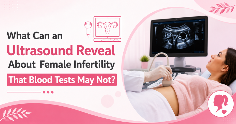
What Can an Ultrasound Reveal About Female Infertility That Blood Tests May Not?
 Deepak
When a woman has trouble conceiving, blood tests are usually the first thing she thinks of. Hormone levels such as AMH, FSH, TSH and prolactin give valuable information. But they show only part of the picture.
Blood tests tell us how the body is functioning chemically. An ultrasound shows us the structure: the uterus, ovaries and surrounding tissues. Many causes of female infertility in Delhi and elsewhere are structural, and a normal blood report does not rule them out.
Why Blood Tests Alone Can Fall Short
A blood test is a snapshot of hormone levels on a particular day. It cannot show whether the uterine lining is healthy, whether a fibroid is pressing on the cavity, or whether a follicle is actually developing and releasing an egg.
This is why many women hear “your reports are normal” yet still struggle to conceive. Normal hormones and a structural problem can exist together.
What an Ultrasound Can Show
A pelvic or transvaginal ultrasound uses sound waves to create real-time images. It is safe, quick and usually painless. Here is what it can reveal.
1. Ovarian Cysts and Polycystic-Appearing Ovaries
Ultrasound can detect cysts, endometriomas (cysts linked to endometriosis) and the multiple small follicles seen in polycystic ovary syndrome (PCOS). A hormone panel may point towards PCOS, but the ovarian appearance on scan adds important detail.
2. Fibroids and Polyps
Fibroids are muscular growths in the uterus, and polyps are small growths in the lining. Depending on their size and position, especially if they distort the uterine cavity, they may interfere with implantation. Blood tests do not detect either.
3. Uterine Shape Differences
Some women are born with a septum (a dividing wall) or other variations in uterine shape. Ultrasound, particularly 3D ultrasound, can suggest these differences. Further tests may be advised to confirm them.
4. Endometrial Thickness and Pattern
The endometrium is the lining where an embryo implants. Ultrasound measures its thickness and appearance at different points in the cycle. A lining that stays too thin, or that looks irregular, may need further evaluation.
5. Ovulation and Follicle Growth
Through follicle monitoring, a doctor can watch a follicle grow across the cycle and confirm whether it ruptures. Some women have regular periods but do not ovulate consistently, and monitoring can help clarify this.
6. Antral Follicle Count
Counting the small follicles visible in the ovaries early in the cycle gives a direct picture of ovarian reserve. Doctors often read this alongside AMH, since each can add to the other.
7. Fluid in the Tubes
A swollen, fluid-filled fallopian tube (hydrosalpinx) can sometimes be seen on ultrasound. This matters because it may reduce the chances of implantation and can affect treatment planning.
8. Adenomyosis and Pelvic Conditions
Features suggesting adenomyosis, where the uterine lining tissue grows into the muscle wall, can sometimes be identified. Ultrasound may also give clues about pelvic adhesions or other conditions that need further assessment.
What Ultrasound Cannot Do
Ultrasound is a strong tool, but it has limits. It cannot confirm whether the fallopian tubes are open, though special tests such as HSG or saline sonography can help with this. It also cannot measure egg quality or show hormone levels.
This is why doctors combine scans, blood tests, medical history and sometimes a semen analysis for the partner. No single test explains everything.
Ultrasound and Blood Tests Work Best Together
QuestionBetter answered byIs ovulation happening?Follicle monitoring and hormone testsHow is ovarian reserve?Antral follicle count with AMHIs the uterus healthy?UltrasoundIs thyroid or prolactin affecting cycles?Blood testsAre there cysts, fibroids or polyps?Ultrasound
When Should You Consider an Evaluation?
Consider speaking with an infertility specialist in Delhi if:
- You are under 35 and have been trying for a year without success
- You are 35 or older and have been trying for six months
- Your periods are irregular, very painful or unusually heavy
- You have a history of pelvic infection, endometriosis, fibroids or ovarian cysts
- You have had miscarriages or a previous pelvic surgery
Earlier assessment is reasonable if any of these apply. Many women wait longer than they need to, simply because their blood tests looked normal.
What to Expect at a Fertility Ultrasound
The scan is usually done in the clinic, often with a transvaginal probe for clearer images. It takes only a few minutes. Timing matters: follicle and lining assessments depend on the cycle day, so your doctor will advise when to come in.
You can bring earlier scans and blood reports. They help your doctor see patterns across time rather than a single day.
Choosing the Right Doctor for Your Evaluation
If you are searching for the best female infertility in Delhi, look beyond the name of a clinic and ask how thorough the evaluation is. A good assessment reads the scan in the context of your history, does not rely on a single report, and explains the findings in plain language.
Dr. Poonam Goyal is a gynecologist and fertility specialist in Delhi who approaches evaluation in this way, combining examination, imaging and laboratory tests to understand each woman’s situation. The aim is a clear plan that suits the individual, not a one-size-fits-all approach. Treatment outcomes depend on many factors, including age, diagnosis and overall health, so no result can be promised.
Key Takeaways
- Blood tests show hormones; ultrasound shows structure.
- Fibroids, polyps, cysts, lining problems and tubal fluid can be missed without a scan.
- Ultrasound has limits and works best alongside other tests.
- A normal report does not always mean there is nothing to find.
If you want consultation regarding female infertility evaluation, you can visit Dr Poonam Goyal at:
- C-3/63A, 64A, opp. Gokulpuri police station, Block C, Panchsheel Hospital, Yamuna Vihar, Shahdara, Delhi, 110053
- HOD IVF & Infertility, Max Superspeciality Hospital, Vaishali
FAQs
1. Can an ultrasound detect infertility?
An ultrasound cannot diagnose infertility on its own, but it can identify structural causes that may contribute to it, such as fibroids, polyps, ovarian cysts or an abnormal uterine lining. It is one part of a complete fertility evaluation.
2. If my blood tests are normal, do I still need an ultrasound?
Often yes. Normal hormone levels do not rule out structural problems in the uterus or ovaries. A scan can add information that blood tests cannot provide.
3. Is a transvaginal ultrasound safe when trying to conceive?
Yes, it is considered safe and is routinely used in fertility assessment. It does not harm a possible pregnancy and is generally well tolerated.
4. Can ultrasound check if my fallopian tubes are blocked?
A standard ultrasound cannot confirm tubal blockage, although it may show fluid in a damaged tube. Tests such as HSG or saline sonography are used to assess tubal patency.
5. When in my cycle should a fertility ultrasound be done?
It depends on what is being checked. Antral follicle count is usually assessed early in the cycle, while follicle growth and lining are tracked around mid-cycle. Your doctor will guide the timing.
Source:
Click for the: Full Story
You might like